VIROLOGY
ISOLATION, CULTIVATION AND IDENTIFICATION OF VIRUSES:
The fact that viruses cannot multiply outside a living host cell
complicates their detection, enumeration, and identification.
Viruses must be provided with living cells instead of a fairly simple
chemical medium. Living plants and animals are difficult and
expensive to maintain. and pathogenic viruses that grow only in
higher primates and human hosts cause additional complications.
However, viruses that usc bacterial cells as a host (bacteriophages)
are rather easily grown on bacterial cultures. This is one
reason so much of our understanding of viral multiplication has
come from bacteriophages.
Growing Bacteriophages in the Laboratory:
Bacteriophages can be grown either in suspensions of bacteria in
liquid media or in bacterial cultures on solid media. The use of
solid media makes possible the plaque method for detecting and
counting viruses. A sample of bacteriophage is mixed with host
bacteria and melted agar. The agar containing the bacteriophages
and host bacteria is then poured into a Petri plate containing a
hardened layer of agar growth medium. The virus-bacteria mixture
solidifies into a thin top layer that contains a layer of bacteria
approximately one cell thick. Each virus infects a bacterium,
multiplies, and releases several hundred new viruses. These
newly produced viruses infect other bacteria in the immediate
vicinity, and more new viruses are produced. Following several
viral multiplication cycles, all the bacteria in the area surrounding
the original virus are destroyed. This produces a number of
clearings, or plaques, visible against a lawn of bacterial growth on the surface of the agar. While the plaques from,
uninfected bacteria elsewhere in the Petri plate multiply rapidly
and produce a turbid background.
Each plaque theoretically corresponds to a single virus in the
initial suspension. Therefore, the concentrations of viral suspensions
measured by the number of plaques are usually given in terms of Plaque Forming Unit(PFU).
 Fig: Plaque formation by bacteriophages
Growing Animal Viruses in the laboratory
In the laboratory, three methods are commonly used for culturing
animal viruses. These methods involve using living animals,
embryonated eggs, or cell cultures.
In Living Animals:
Some animal viruses can be cultured only in living animals, such
as mice, rabbits, and guinea pigs. Most experiments to study the
immune system's response to viral infections must also be performed
in virally infected live animals. Animal inoculation may
be used as a diagnostic procedure for identifying and isolating a
virus from a clinical specimen. After the animal is inoculated
with the specimen, the animal is observed for signs of disease or
is killed so that infected tissues can be examined for the virus.
Some human viruses cannot be grown in animals or can be
grown but do not cause disease. The lack of natural animal models for AIDS has slowed our understanding of its disease
process and prevented experimentation with drugs that inhibit
growth of the virus in vivo. Chimpanzees can be infected with
one subspecies of human immunodeficiency virus (HIV- l, genus
Lentivirus ), but because they do not show symptoms of the disease,
they cannot be used to study the effects of viral growth and
disease treatments. AIDS vaccines are presently being tested in
humans, but the disease progresses so slowly in humans that it
can take years to determine the effectiveness of these vaccines. In
1986, simian AIDS (an immunodeficiency disease of green monkeys)
was reported, followed in 1987 by feline AIDS (an immunodeficiency
disease of domestic cats). These diseases are caused by lentiviruses, which are closely related to HIV, and the diseases develop within a few months, thus providing model for studying viral growth in different tissues.
In 1990, a way to infect mice
with human AIDS was found when immunodeficient mice were
grafted to produce human T cells and human gamma globulin.
The mice provide a reliable model for studying viral replication,
although they do not provide models for vaccine development.
In Embryonated Eggs:
If the virus will grow in an embryonated egg, this can be a fairly
convenient and inexpensive form of host for many animal viruses.
A hole is drilled in the shell of the embryonated egg, and a viral
suspension or suspected virus-containing tissue is injected into the
fluid of the egg. There are several membranes in an egg, and the
virus is injected near the one most appropriate for its growth. Viral growth is signaled by the death of the embryo, by embryo cell damage, or by the formation of typical pocks or
lesions on the egg membranes. This method was once the most
widely used method of viral isolation and growth, and it is still
used to grow viruses for some vaccines. For this reason, you may
be asked if you are allergic to eggs before receiving a vaccination,
because egg proteins may be present in the viral vaccine preparations.
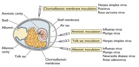 Fig: Routes of growing viruses in embryonic egg
In Cell Cultures:
Cell cultures have replaced embryonated eggs as the preferred
type of growth medium for many vi ruses. Cell cultures consist of
cells grown in culture media in the laboratory. Because these cultures
are generally rather homogeneous collections of cells and
can be propagated and handled much like bacterial cultures, they
are more convenient to work with than whole animals or embryonated eggs.
Cell culture lines are started by treating a slice
 of animal tissue
with enzymes that separate the individual cells .
These cells are suspended in a solution that provides the osmotic
pressure, nutrients, and growth factors needed for the cells to
grow. Normal cells tend to adhere to the glass or plastic container
and reproduce to form a monolayer. Viruses infecting such a
monolayer sometimes cause the cells of the monolayer to deteriorate
as they multiply. This cell deterioration is called cytopathic
effect (CPE) . CPE can be detected and
counted in much the same way as plaques caused by bacteriophages
on a lawn of bacteria and reported as PFU /mL
Viruses may be grown in primary or continuous cell lines.
Primary cell lines, derived from tissue slices, tend to die out
after only a few generations. Certain cell lines, called diploid cell
lines, developed from human embryos can be maintained for
about 100 generations and are widely used for culturing viruses
that require a human host. Cell lines developed from embryonic
human cells are used to culture rabies virus for a rabies vaccine
called human diploid culture vaccine .
When viruses are routinely grown in a laboratory, continuous
cell lines are used. These are transformed (cancerous) cells that
can be maintained through an indefinite number of generations,
and they are sometimes called immortal cell lines . One of these, the HeLa cell
line, was isolated from the cancer of a woman (Henrietta Lacks)
who died in 1951. After years of laboratory cultivation, many such
cell lines have lost almost all the original characteristics of the cell,
but these changes have not interfered with the use of the cells for
viral propagation. In spite of the success of cell culture in viral
isolation and growth, there are still some viruses that have never
been successfully cultivated in cell culture.
The idea of cell culture dates back to the end of the nineteenth
century, but it was not a practical laboratory technique.


Cited By Kamal Singh Khadka
Msc Microbiology, TU
The fact that viruses cannot multiply outside a living host cell
complicates their detection, enumeration, and identification.
Viruses must be provided with living cells instead of a fairly simple
chemical medium. Living plants and animals are difficult and
expensive to maintain. and pathogenic viruses that grow only in
higher primates and human hosts cause additional complications.
However, viruses that usc bacterial cells as a host (bacteriophages)
are rather easily grown on bacterial cultures. This is one
reason so much of our understanding of viral multiplication has
come from bacteriophages.
Growing Bacteriophages in the Laboratory:
Bacteriophages can be grown either in suspensions of bacteria in
liquid media or in bacterial cultures on solid media. The use of
solid media makes possible the plaque method for detecting and
counting viruses. A sample of bacteriophage is mixed with host
bacteria and melted agar. The agar containing the bacteriophages
and host bacteria is then poured into a Petri plate containing a
hardened layer of agar growth medium. The virus-bacteria mixture
solidifies into a thin top layer that contains a layer of bacteria
approximately one cell thick. Each virus infects a bacterium,
multiplies, and releases several hundred new viruses. These
newly produced viruses infect other bacteria in the immediate
vicinity, and more new viruses are produced. Following several
viral multiplication cycles, all the bacteria in the area surrounding
the original virus are destroyed. This produces a number of
clearings, or plaques, visible against a lawn of bacterial growth on the surface of the agar. While the plaques from,
uninfected bacteria elsewhere in the Petri plate multiply rapidly
and produce a turbid background.
Each plaque theoretically corresponds to a single virus in the
initial suspension. Therefore, the concentrations of viral suspensions
measured by the number of plaques are usually given in terms of Plaque Forming Unit(PFU).
Growing Animal Viruses in the laboratory
In the laboratory, three methods are commonly used for culturing
animal viruses. These methods involve using living animals,
embryonated eggs, or cell cultures.
In Living Animals:
Some animal viruses can be cultured only in living animals, such
as mice, rabbits, and guinea pigs. Most experiments to study the
immune system's response to viral infections must also be performed
in virally infected live animals. Animal inoculation may
be used as a diagnostic procedure for identifying and isolating a
virus from a clinical specimen. After the animal is inoculated
with the specimen, the animal is observed for signs of disease or
is killed so that infected tissues can be examined for the virus.
Some human viruses cannot be grown in animals or can be
grown but do not cause disease. The lack of natural animal models for AIDS has slowed our understanding of its disease
process and prevented experimentation with drugs that inhibit
growth of the virus in vivo. Chimpanzees can be infected with
one subspecies of human immunodeficiency virus (HIV- l, genus
Lentivirus ), but because they do not show symptoms of the disease,
they cannot be used to study the effects of viral growth and
disease treatments. AIDS vaccines are presently being tested in
humans, but the disease progresses so slowly in humans that it
can take years to determine the effectiveness of these vaccines. In
1986, simian AIDS (an immunodeficiency disease of green monkeys)
was reported, followed in 1987 by feline AIDS (an immunodeficiency
disease of domestic cats). These diseases are caused by lentiviruses, which are closely related to HIV, and the diseases develop within a few months, thus providing model for studying viral growth in different tissues.
In 1990, a way to infect mice
with human AIDS was found when immunodeficient mice were
grafted to produce human T cells and human gamma globulin.
The mice provide a reliable model for studying viral replication,
although they do not provide models for vaccine development.
In Embryonated Eggs:
If the virus will grow in an embryonated egg, this can be a fairly
convenient and inexpensive form of host for many animal viruses.
A hole is drilled in the shell of the embryonated egg, and a viral
suspension or suspected virus-containing tissue is injected into the
fluid of the egg. There are several membranes in an egg, and the
virus is injected near the one most appropriate for its growth. Viral growth is signaled by the death of the embryo, by embryo cell damage, or by the formation of typical pocks or
lesions on the egg membranes. This method was once the most
widely used method of viral isolation and growth, and it is still
used to grow viruses for some vaccines. For this reason, you may
be asked if you are allergic to eggs before receiving a vaccination,
because egg proteins may be present in the viral vaccine preparations.
In Cell Cultures:
Cell cultures have replaced embryonated eggs as the preferred
type of growth medium for many vi ruses. Cell cultures consist of
cells grown in culture media in the laboratory. Because these cultures
are generally rather homogeneous collections of cells and
can be propagated and handled much like bacterial cultures, they
are more convenient to work with than whole animals or embryonated eggs.
Cell culture lines are started by treating a slice
with enzymes that separate the individual cells .
These cells are suspended in a solution that provides the osmotic
pressure, nutrients, and growth factors needed for the cells to
grow. Normal cells tend to adhere to the glass or plastic container
and reproduce to form a monolayer. Viruses infecting such a
monolayer sometimes cause the cells of the monolayer to deteriorate
as they multiply. This cell deterioration is called cytopathic
effect (CPE) . CPE can be detected and
counted in much the same way as plaques caused by bacteriophages
on a lawn of bacteria and reported as PFU /mL
Viruses may be grown in primary or continuous cell lines.
Primary cell lines, derived from tissue slices, tend to die out
after only a few generations. Certain cell lines, called diploid cell
lines, developed from human embryos can be maintained for
about 100 generations and are widely used for culturing viruses
that require a human host. Cell lines developed from embryonic
human cells are used to culture rabies virus for a rabies vaccine
called human diploid culture vaccine .
When viruses are routinely grown in a laboratory, continuous
cell lines are used. These are transformed (cancerous) cells that
can be maintained through an indefinite number of generations,
and they are sometimes called immortal cell lines . One of these, the HeLa cell
line, was isolated from the cancer of a woman (Henrietta Lacks)
who died in 1951. After years of laboratory cultivation, many such
cell lines have lost almost all the original characteristics of the cell,
but these changes have not interfered with the use of the cells for
viral propagation. In spite of the success of cell culture in viral
isolation and growth, there are still some viruses that have never
been successfully cultivated in cell culture.
The idea of cell culture dates back to the end of the nineteenth
century, but it was not a practical laboratory technique.
Cited By Kamal Singh Khadka
Msc Microbiology, TU

Comments