Posts

Showing posts from February, 2013

MICROBIAL METABOLISM CONTD...

Image
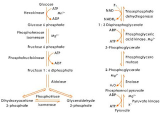
GLYCOLYSIS: Steps:       The Embden-Meyerhof or glycolytic pathway is undoubtedly the most common pathway for glucose degradation to pyruvate in stage two of catabolism. It is found in all major groups of microorganisms and functions in the presence or absence of O2. Glycolysis [Greek glyco, sweet, and lysis, a loosening] is located in the cytoplasmic matrix of procaryotes and eucaryotes. The pathway as a whole may be divided into two parts.  In the initial six-carbon stage, glucose is phosphorylated twice and eventually converted to fructose 1,6- bisphosphate. Other sugars are often fed into the pathway by conversion to glucose 6-phosphate or fructose 6-phosphate. This preliminary stage does not yield energy; in fact, two ATP molecules are expended for each glucose. These initial steps “prime the pump” by adding phosphates to each end of the sugar. The phosphates will soon be used to make ATP. The three-carbon stage of glycolysis begins when the enzyme fructose 1,

MICROBIAL METABOLISM

Image
Living cells are in a state of ceaseless activity. To maintain its “life,” each cell depends on highly coordinated biochemical reactions. Carbohydrates are an important source of the energy that drives these reactions. This chapter discusses the energy-generating pathways of carbohydrate metabolism are discussed. During glycolysis, an ancient pathway found in almost all organisms, a small amount of energy is captured as a glucose molecule is converted to two molecules of pyruvate. Glycogen, a storage form of glucose in vertebrates, is synthesized by glycogenesis when glucose levels are high and degraded by glycogenolysis when glucose is in short supply. Glucose can also be synthesized from noncarbohydrate precursors by reactions referred to as gluconeogenesis. The pentose phosphate pathway enables cells to convert glucose-6-phosphate, a derivative of glucose, to ribose- 5-phosphate (the sugar used to synthesize nucleotides and nucleic acids) and other types of monosacchar

MICROBIAL METABOLISM

Image
Metabolism:  Metabolism is the dynamic balance between difference types of reaction( catabolism+ anabolism) that provide the cell with energy or building materials & those to utilize them. It consists of two general activities:  i)  Energy Production                             ii) Energy Utilization  Metabolic pathways are treated as a sequence of enzymes functioning as a unit, with each enzyme using as its substrate a product of the preceding enzyme-catalyzed reaction. This picture of metabolic pathways is incomplete because we will usually ignore the regulation of pathway operation for the sake of space and simplicity. However, one should keep in mind that both regulation of the activity of individual pathways and coordination of the action of separate sequences are essential to the existence of life. Cells become disorganized and die without adequate control of metabolism, and regulation is just as important to life as is the efficient use of energy. Thus the last

BACTERIAL MORPHOLOGY CONTD..

Image
CELL STRUCTURE INTERNAL TO CELL WALL: 3). Inclusion Bodies:   There are granules of organic or inorganic materials. These bodies are generally for storage( Eg  carbon compound, inorganic substance & energy) & also helps to osmotic pressure. Some of the inclusion bodies are poly-beta- hydroxy butyrate  granules( carbon storage), some glucose(carbon storage), sulphur granules( sulphur storage), gas vacuoles( helps bacteria to regulate buoyancy & float), phosphate granules or volutin granules or meta chromatin granules( storage of phosphates). Mesosomes:  The cytoplasmic membrane is invaginated  at certain places into the cytoplasm in form of convoluted tubules  & vesicles known as mesosomes. On these surfaces are found enzymes associated to mitochondria of eucaryotic. Most of mesosomes are confined to the periphery showing only a shallow penetration.However, some penetrate deeply into the cytoplasm near to the cell's nuclear material. These  are thought to be in

BACTERIAL MORPHOLOGY CONTD...

STRUCTURE INTERNAL TO CELL WALL: 2.) Cytoplasm :  It refers to internal matrix of cell inside the cytoplasmic membrane. Prokaryotic cytoplasm lacks membrane bound structures . The cytoplasmic matrix is largely watered which dissolves proteins, amino acids, sugars, nucleotides, salts,  vitamins, & ions. It also contains certain bacterial structures like inclusion bodies, nucleoid, plasmids. A) Ribosomes :   They are r-RNA protein bodies which give granular appearance.  Proteins  are synthesized in ribosomes & prokaryotic. Proteins are synthesized  in ribosomes & prokaryotic ribosomes aren't bound to endoplasmic  reticulum. Ribosomes of prokaryotes are called 70S(Sedimentation Coefficient) & are composed of 2 units(50S & 30S). Antibiotics such as Streptomycin &Tetracycline prevents bacterial ribosomes from carrying out protein synthesis. B). Nucleoid:    It is an irregular shape region where prokaryotic chromosomes are located. The nucleoid doesn'

BACTERIAL MORPHOLOGY CONTD...

Image
B) STRUCTURE INTERNAL TO CELL WALL; 1) Cytoplasmic membrane:   It is a thick structure( 8 nm)  that separates the cytoplasm from outer membrane. It is composed of phospholipid & protein. Phospholipid forms the bi layer in which protein( integral & peripheral) are held. Functions: It acts as permeability barrier & prevents the leakage of cytoplasmic constituents  into or out of the cell.  Specific proteins in membrane facilitates passage of small molecules across the membrane. It contains several enzymes involved in respiratory metabolism & in synthesis of cell wall & capsular components.  Note:  Polymyxins  makes holes on cytoplasmic membrane while detergents & alcohol dissolves the bi-layers.

BACTERIAL MORPHOLOGY CONTD...

Image
A) STRUCTURE EXTERNAL TO CELL WALL 4)  Cell Wall:  1) Gram Positive Cell Wall  : Gram Positive cell wall has greater amount of peptidoglycan (ie around 25 nanometer) & may account for 50% or more of total dry weight of cell. Besides, peptidoglycan gram positive cell wall contains Teichoic acid. It helps to link the peptidoglycan layer & provides negative charge to the cell wall. Though, much gram positive cell wall contains very little lipid but Mycobacteria &  Cornyebacterium spp   & other are rich in lipids called Mycolic acid.                                                       FIG:  B= GRAM NEGATIVE CELL WALL A= GRAM POSITIVE CELL WALL                                                             2). Gram Negative Cell Wall:   Wall of Gram negative bacteria are more complex than those of Gram positive bacteria. It comprises lesser amount of peptidoglycan (2 nm to 3 nm) thick,which  accounts 10% of wall weight. The outer membrane is absent in Gram posi

BACTERIAL MORPHOLOGY CONTD...

Image
STRUCTURE EXTERNAL TO CELL WALL 4) Cell wall:   It is a rigid structure that lies beneath external structures( Pili, capsule, flagella) & external to cytoplasmic membrane. It gives shape & rigidity  to cell. Cell wall consists network of peptidoglycan layer( murein or mucopeptide). Structure Of Peptidoglycan layer:                       A peptidoglycan is only found in prokaryotic cell surrounding cytoplasm & differs  from species to species. Basically, it is composed alternating unit of NAM(N-acteylmuramic acid) & NAG(N-acteylglucosamine). It also consists of peptide of four or five amino acids ( L-alanine, D-alanine , D-glutamate,  and either lysine or Diaminopimelic acid(DAP).                    So, peptidoglycan simply  consists of polysaccharides backbone composed of alternating units of NAG & NAM  with short peptide chain projecting from NAM units. There peptide chains are cross linked with each other & this cross linkage can be different  in Gr

BACTERIAL MORPHOLOGY CONTD...

Image
STRUCTURE EXTERNAL TO CELL WALL : 2).   Pili or Fimbriae  :   They are thin,soft, hair like appendages that extends from cytoplasmic membrane of Gram negative bacteria . They are shorter, straighter & numerous than flagella. They are composed of pilin protein, but don't function as motility. Advantages:                                                        Fig: Prokaryotic cell structure showing Pili. fig:   Schematic drawing of bacterial conjugation.  1-  Donor cell produces pilus.  2-  Pilus attaches to recipient cell, brings the two cells together.  3- The mobile plasmid is nicked and a single strand of DNA is then transferred to the recipient cell.  4-  Both cells recircularize their plasmids, synthesize second strands, and reproduce pili; both cells are now viable donors. Helps in attachment ie allows pathogenic bacteria to attach epithelial cells, linings of respiratory tract, intestinal tract etc. So, Pili  helps in colonization & prevents bacter

BACTERIAL MORPHOLOGY CONTD....

Image
STRUCTURE OF BACTERIAL CELL WALL A). Structure External To Cell Wall : 1). Flagella  : They are hair like appendages that protrudes from cell wall & are responsible for motility. Each of flagella is composed of filament, hook,& basal body.                                                  fig : Ultra structure Of Bacterium Flagella    Filaments is attached to hook like structure & composed of flagellin protein . Hook lies at the base  of flagella & filament connects to basal body. Basal body forms a central rod that consists of set of enclosing rings. Gram positive bacteria has only one ring embedded in cell membrane & one in cell wall while gram negative bacteria have a pair of rings embedded in cell membrane & another pair in cell wall.  According to arrangement of flagella, bacteria can be classified as;                       a) Monotrichous :  Having only one flagella. Eg: Vibrio cholerae , Pseudomonas                         b)

BACTERIAL MORPHOLOGY Contd...

Image
SHAPE & ARRANGEMENT c). Spirals:  They are spiral shaped bacteria. Some are curved rods that resembles comma. Eg: Vibrio cholerae .    Spirilla:  Helical shape with a thick, rigid cell wall & flagella that resist in movement. Eg: Spirillum spp  .  Spirochetes: Spiral shaped bacteria with a thin, flexible cell wall but as flagella. Eg: Treponema palladium.                                           fig:  Vibrio cholerae                                                                                                 fig: Treponema   pallidum     at high resolution                                                                                          Fig:  Vibrio cholerae                                                   fig:  Treponema  pallidum                                             fig: Spirillum spp

BACTERIAL MORPHOLOGY Contd..

Image
SHAPE & ARRANGEMENT(Contd...) b). Bacilli or Rods :  Bacilli are rods shaped bacteria. Some are slender( Salmonella typhi ), some are rectangular with square ends( Bacillus anthracis ), some are cluster shaped( Clostridium diptheriae ).  Bacilli aren't arranged as complex as that of cocci, however the arrangement are diplobacilli(cells arranged in pairs), Lactobacillius spp , Streptobacillus spp , cells arranged in chain, trichomes(similar to chains,but have  larger area of contact between adjacent cells). Baggiota spp i). Palisades:  cells are lined side by side like match sticks & at angles to one another.  Eg . Cornyebacterium spp.                                            Fig:     Salmonella typhi                                                                                                               fig. Lactobacillus spp                                              fig. Streptobacillus spp