VIROLOGY
Host Range:
The host range of a virus is the spectrum of host cells the virus can
infect. There are viruses that infect invertebrates, vertebrates,
plants, protists, fungi, and bacteria. However, most viruses are able
to infect specific types of cells of only one host species. In rare
cases, viruses cross the host-range barrier, thus expanding their
host range. Viruses that infect bacteria are called
bacteriophages, or phages.
The particular host range of a virus is determined by the
virus's requirements for its specific attachment to the host cell
and the availability within the potential host of cellular factors
required for viral multiplication. For the virus to infect the host
cell, the outer surface of the virus must chemically interact
with specific receptor sites on the surface of the celL The two
complementary components are held together by weak bonds,
such as hydrogen bonds. The combination of many attachment
and receptor sites leads to a strong association between host cell
and virus. For some bacteriophages, the receptor site is part of
the cell wall of the host; in other cases, it is part of the fimbriae
or flagella. For animal viruses, the receptor sites are on the plasma
membranes of the host cells.
The potential to use viruses to treat diseases is intriguing
because of their narrow host range and their ability to kill their host cells. The idea of phage therapy----using bacteriophage to
treat bacterial infections, has been around for 100 years. Recent
advances in OUT understanding of virus-host interactions have
fueled new studies in the field of phage therapy.
Experimentally induced viral infections in cancer patients
during the 1920s suggested that viruses might have antitumor
activity. These tumor-destroying, or oncolytic, viruses may selectively
infect and kill tumor cells or cause an immune response
against tumor cells. Some viruses naturally infect tumor cells,
and other viruses can be genetically modified to infect tumor
cells. At present several studies arc underway to determine the
killing mechanism of oncolytic viruses and the safety of using viral therapy.
VIRAL SIZE:
Viral sizes are determined with the aid of electron microscopy.
Different viruses vary considerably in size. Although most are
quite a bit smaller than bacteria, some of the larger viruses (such
as the vaccinia virus) are about the same size as some very small
bacteria (such as the mycoplasmas, rickettsias, and chlamydias).
Viruses range from 20 to 1000 nm in length.
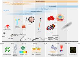
Fig: Comparison of viral size
Viral Structure:
A virion is a complete, fully developed, infectious viral particle
composed of nucleic acid and surrounded by a protein coat that
protects it from the environment and is a vehicle of transmission
from one host cell to another. Viruses are classified by differences
in the structures of these coats.
Nucleic Acid:
In contrast to prokaryotic and eukaryotic cells, in which DNA is
always the primary genetic material (and RNA plays an auxiliary
role), a virus can have either DNA or RNA- but never both. The
nucleic acid of a virus can be single-stranded or double-stranded.
Thus, there are viruses with the familiar double-stranded DNA,
with single-stranded DNA, with double-stranded RNA, and with
single-stranded RNA. Depending on the virus, the nucleic acid can be linear or circular. In some viruses (such as the influenza
virus), the nucleic acid is in several separate segments.
The percentage of nucleic acid in relation to protein is about
1 % for the influenza virus and about 50% for certain bacteriophages.
The total amount of nucleic acid varies from a few thousand
nucleotides (or pairs) to as many as 250,000 nucleotides.
( E. coli chromosome consists of approximately 4 million nucleotide pairs).

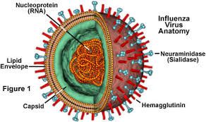
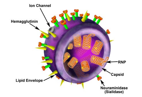
Capsid and Envelope:
The nucleic acid of a virus is protected by a protein coat called
the capsid . The structure of the capsid is ultimately
determined by the viral nucleic acid and accounts for
most of the mass of a virus, especially of small ones. Each capsid
is composed of protein subunits called capsomeres. In some
viruses, the proteins composing the capsomeres are of a single
type; in other viruses, several types of protein may be present.
Individual capsomeres are often visible in electron micrographs.The arrangement of capsomeres
is characteristic of a particular type of virus .
In some viruses, the capsid is covered by an envelope
, which usually consists of some combination of
lipids, proteins, and carbohydrates. Some animal viruses are
released from the host cell by an extrusion process that coats
the virus with a layer of the host cell's plasma membrane; that
layer becomes the viral envelope. In many cases, the envelope
contains proteins determined by the viral nucleic acid and
materials derived from normal host cell components.
Depending on the virus, envelopes mayor may not be covered
by spikes, which are carbohydrate-protein complexes that
project from the surface of the envelope. Some viruses attach to
host cells by means of spikes. Spikes are such a reliable characteristic
of some viruses that they can be used as a means of identification.
The ability of certain viruses, such as the influenza virus
, to clump red blood cells is associated with spikes.
Such viruses bind to red blood cells and form bridges between them. The resulting clumping is called haemagglutination and is the basis for several laboratory tests.
Cited by My Beloved Teacher
Kamal Singh Khadka
Msc Microbiology, TU
The host range of a virus is the spectrum of host cells the virus can
infect. There are viruses that infect invertebrates, vertebrates,
plants, protists, fungi, and bacteria. However, most viruses are able
to infect specific types of cells of only one host species. In rare
cases, viruses cross the host-range barrier, thus expanding their
host range. Viruses that infect bacteria are called
bacteriophages, or phages.
The particular host range of a virus is determined by the
virus's requirements for its specific attachment to the host cell
and the availability within the potential host of cellular factors
required for viral multiplication. For the virus to infect the host
cell, the outer surface of the virus must chemically interact
with specific receptor sites on the surface of the celL The two
complementary components are held together by weak bonds,
such as hydrogen bonds. The combination of many attachment
and receptor sites leads to a strong association between host cell
and virus. For some bacteriophages, the receptor site is part of
the cell wall of the host; in other cases, it is part of the fimbriae
or flagella. For animal viruses, the receptor sites are on the plasma
membranes of the host cells.
The potential to use viruses to treat diseases is intriguing
because of their narrow host range and their ability to kill their host cells. The idea of phage therapy----using bacteriophage to
treat bacterial infections, has been around for 100 years. Recent
advances in OUT understanding of virus-host interactions have
fueled new studies in the field of phage therapy.
Experimentally induced viral infections in cancer patients
during the 1920s suggested that viruses might have antitumor
activity. These tumor-destroying, or oncolytic, viruses may selectively
infect and kill tumor cells or cause an immune response
against tumor cells. Some viruses naturally infect tumor cells,
and other viruses can be genetically modified to infect tumor
cells. At present several studies arc underway to determine the
killing mechanism of oncolytic viruses and the safety of using viral therapy.
VIRAL SIZE:
Viral sizes are determined with the aid of electron microscopy.
Different viruses vary considerably in size. Although most are
quite a bit smaller than bacteria, some of the larger viruses (such
as the vaccinia virus) are about the same size as some very small
bacteria (such as the mycoplasmas, rickettsias, and chlamydias).
Viruses range from 20 to 1000 nm in length.
Fig: Comparison of viral size
Viral Structure:
A virion is a complete, fully developed, infectious viral particle
composed of nucleic acid and surrounded by a protein coat that
protects it from the environment and is a vehicle of transmission
from one host cell to another. Viruses are classified by differences
in the structures of these coats.
Nucleic Acid:
In contrast to prokaryotic and eukaryotic cells, in which DNA is
always the primary genetic material (and RNA plays an auxiliary
role), a virus can have either DNA or RNA- but never both. The
nucleic acid of a virus can be single-stranded or double-stranded.
Thus, there are viruses with the familiar double-stranded DNA,
with single-stranded DNA, with double-stranded RNA, and with
single-stranded RNA. Depending on the virus, the nucleic acid can be linear or circular. In some viruses (such as the influenza
virus), the nucleic acid is in several separate segments.
The percentage of nucleic acid in relation to protein is about
1 % for the influenza virus and about 50% for certain bacteriophages.
The total amount of nucleic acid varies from a few thousand
nucleotides (or pairs) to as many as 250,000 nucleotides.
( E. coli chromosome consists of approximately 4 million nucleotide pairs).
Capsid and Envelope:
The nucleic acid of a virus is protected by a protein coat called
the capsid . The structure of the capsid is ultimately
determined by the viral nucleic acid and accounts for
most of the mass of a virus, especially of small ones. Each capsid
is composed of protein subunits called capsomeres. In some
viruses, the proteins composing the capsomeres are of a single
type; in other viruses, several types of protein may be present.
Individual capsomeres are often visible in electron micrographs.The arrangement of capsomeres
is characteristic of a particular type of virus .
In some viruses, the capsid is covered by an envelope
, which usually consists of some combination of
lipids, proteins, and carbohydrates. Some animal viruses are
released from the host cell by an extrusion process that coats
the virus with a layer of the host cell's plasma membrane; that
layer becomes the viral envelope. In many cases, the envelope
contains proteins determined by the viral nucleic acid and
materials derived from normal host cell components.
Depending on the virus, envelopes mayor may not be covered
by spikes, which are carbohydrate-protein complexes that
project from the surface of the envelope. Some viruses attach to
host cells by means of spikes. Spikes are such a reliable characteristic
of some viruses that they can be used as a means of identification.
The ability of certain viruses, such as the influenza virus
, to clump red blood cells is associated with spikes.
Such viruses bind to red blood cells and form bridges between them. The resulting clumping is called haemagglutination and is the basis for several laboratory tests.
Cited by My Beloved Teacher
Kamal Singh Khadka
Msc Microbiology, TU

Comments