The Study of Microbial Structure: Microscopy and Specimen Preparation contd..
Types Of Electron Microscope
2. Scanning Electron Microscope
Here, the Specimen is subjected to a narrow electron beam which rapidly moves over the surface of it. This causes the release of secondary electrons which are corrected by a detector that generates an electronic signal. This signal are then scanned to produce the image. This electron microscope is used for visualizing three dimensional pictures(3D) .
The previously described microscopes form an image from radiation
that has passed through a specimen. More recently the
scanning electron microscope (SEM) has been used to examine
the surfaces of microorganisms in great detail; many instruments
have a resolution of 7 nm or less. The SEM differs from
other electron microscopes in producing an image from electrons
emitted by an object’s surface rather than from transmitted
electrons.
Specimen preparation is easy, and in some cases air-dried
material can be examined directly. Most often, however, microorganisms
must first be fixed, dehydrated, and dried to preserve
surface structure and prevent collapse of the cells when they
are exposed to the SEM’s high vacuum. Before viewing, dried
samples are mounted and coated with a thin layer of metal to prevent
the buildup of an electrical charge on the surface and to give
a better image.
The SEM scans a narrow, tapered electron beam back and
forth over the specimen. When the beam strikes a
particular area, surface atoms discharge a tiny shower of electrons
called secondary electrons, and these are trapped by a special detector.
Secondary electrons entering the detector strike a scintillator
causing it to emit light flashes that a photo multiplier converts
to an electrical current and amplifies. The signal is sent to a
cathode-ray tube and produces an image like a television picture which can be viewed or photographed.
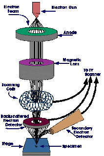
Fig: Working of SEM

Comments