VIROLOGY
MULTIPLICATION OF ANIMAL VIRUSES:
The multiplication of animal viruses follows the basic pattern
of bacteriophage multiplication but has several differences.
Animal viruses differ from phages in
their mechanism of entering the host cell. Also, once the virus is
inside, the synthesis and assembly of the new viral components
are somewhat different, partly because of the differences between
prokaryotic cells and eukaryotic cells. Animal viruses may have
certain types of enzymes not found in phages. Finally, the mechanisms
of maturation and release, and the effects on the host cell,
differ in animal viruses and phages.
In the following discussion of the multiplication of animal
viruses, we will consider the processes that are shared by both
DNA- and RNA-containing animal viruses. These processes are
attachment, entry, uncoating, and release. We will also examine
how DNA- and RNA-containing viruses differ with respect to their process of biosynthesis.

Attachment: Like bacteriophages, animal viruses have attachment sites that
attach to complementary receptor sites on the host cell's surface.
However, the receptor sites of animal cells are proteins
and glycoproteins of the plasma membrane. Moreover, animal
viruses do not possess appendages like the tail fibers of some
bacteriophages. The attachment sites of animal viruses are distributed
over the surface of the virus. The sites themselves vary
from one group of viruses to another. In adenoviruses, which
are icosahedral viruses, the attachment sites are small fibers at
the corners of the icosahedron . In many of
the enveloped viruses, such as influenza virus, the attachment
sites are spikes located on the surface of the envelope . As soon as one spike attaches to a host receptor,
additional receptor sites on the same cell migrate to the virus.
Attachment is completed when many sites are bound.
Receptor sites arc inherited characteristics of the host. Receptor sites arc inherited characteristics of the host.
Consequently, the receptor for a particular virus can vary from
person to person. This could account for the individual differences
in susceptibility to a particular virus. For example, people
who lack the cellular receptor (called P antigen) for parvovirus
B19, are naturally resistant to infection and do not get fifth disease.
Understanding the nature of attachment can
lead to the development of drugs that prevent viral infections.
Monoclonal antibodies that combine
with a virus's attachment site or the cell's receptor site may soon be used to treat some viral infections.
Entry:
Following attachment, entry occurs. Viruses enter into
eukaryotic cells by pinocytosis, an active cellular process by
which nutrients and other molecules are brought into a cell.
A cell's plasma membrane continuously
folds inward to form vesicles. These vesicles contain elements
that originate outside the cell and are brought into the
interior of the cell to be digested . If a virion attaches to the
plasma membrane of a potential host cell, the host cell will enfold the virion into a fold of plasma membrane, forming a vesicle.
Enveloped viruses can enter by an alternative method called
fusion, in which the viral envelope fuses with the plasma membrane
and releases the capsid into the cell's cytoplasm. For example,
HIV penetrates cells by this method .
Uncoating:
Viruses disappear during the eclipse period of an infection
because they are taken apart inside the cell. Uncoating is the separation
of the viral nucleic acid from its protein coat once the
virion is enclosed within the vesicle. The capsid is digested when
the cell attempts to digest the vesicle's contents, or the non enveloped
capsid may be released into the cytoplasm of the host cell.
This process varies with the type of virus. Some animal viruses
accomplish uncoating by the action of lysosomal enzymes of the
host cell. These enzymes degrade the proteins of the viral capsid.
The uncoating of poxviruses is completed by a specific enzyme
encoded by the viral DNA and synthesized soon after infection.
For other viruses, uncoating appears to be exclusively caused by
enzymes in the host cell cytoplasm. For at least one virus, the poliovirus, uncoating seems 10 begin while the virus attached to the host cell's plasma membrane.
The Biosynthesis of DNA Viruses:
Generally, DNA-containing viruses replicate their DNA in the
nucleus of the host cell by using viral enzymes, and they synthesize
their capsid and other proteins in the cytoplasm by
using host cell enzymes. Then the proteins migrate into the
nucleus and are joined with the newly synthesized DNA to
form virions. These virions are transported along the endoplasmic
reticulum to the host cell's membrane for release.
Herpesviruses, papovaviruses, adenoviruses s, and hepadnaviruses
all follow this pattern of biosynthesis.
Poxviruses are an exception because all of their components are synthesized in cytoplasm.


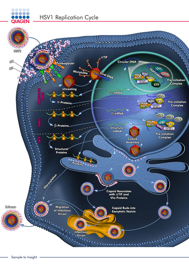


Fig: Viral Replication (Animal Viruses Life Cycle)
Viruses and Cancer :
Several types of cancer are now known to be caused by viruses.
Molecular biological research shows that the mechanisms of the
diseases are similar, even when a virus does not cause the cancer.
The relationship between cancers and viruses was first
demonstrated in 1908, when virologists Wilhelm Ellerman and
Olaf Bang, working in Denmark, were trying to isolate the
causative agent of chicken leukemia. They found that leukemia
could be transferred to healthy chickens by cell-free filtrates that
contained viruses. Three years later, F. Peyton Rous, working at
the Rockefeller Institute in New York, found that a chicken
sarcoma (cancer of connective tissue) can be similarly transmitted
. Virus-induced adenocarcinomas (ca ncers of glandular
epithelial tissue) in mice were discovered in 1936.At that time, it
was clearly shown that mouse mammary gland tumors are transmitted
from mother to offspring through the mother's milk.
A human cancer-causing virus was discovered and isolated in
1972 by American bacteriologist Sarah Stewart.
The viral cause of cancer can often go unrecognized for several
reasons. First, most of the particles of some viruses infect
cells but do not induce cancer. Second, cancer might not develop
until long after viral infection. Third, cancers do not seem to be
contagious, as viral diseases usually are.
The Transformation of Normal Cells Into Tumor Cells:
Almost anything that can alter the genetic material of a eukaryotic
cell has the potential to make a normal cell cancerous. These
cancer-causing alterations to cellular DNA affect parts of the
genome called oncogenes. Oncogenes were first identified in cancer-causing viruses and were thought to be a part of the normal
viral genome. However, American microbiologists J. Michael
Bishop and Harold E. Varmus received the 1989 Nobel Prize in
Medicine for proving that the cancer-inducing genes carried by
viruses are actually derived from animal cells. Bishop and E.Varmus
showed that the cancer-causing src gene in avian sarcoma
viruses is derived from a normal part of chicken genes.
Oncogenes can be activated to abnormal functioning by a
variety of agents, including mutagenic chemicals, high -energy
radiation, and viruses. Viruses capable of inducing tumors in
animals are called oncogenic viruses, or oncoviruses.
Approximately 10% of cancers are known to be virus-induced.
An outstanding feature of all oncogenic viruses is that their
genetic material integrates into the host cell's DNA and replicates
along with the host cell's chromosome. This mechanism is similar
to the phenomenon of lysogeny in bacteria, and it can alter
the host cell's characteristics in the same way.
Tumor cells undergo transformation; that is, they acquire
properties that are distinct from the properties of uninfected
cells or from infected cells that do not form tumors. After being
transformed by viruses, many tumor cells contain a virus specific
antigen on their cell surface, called tumor-specific
transplantation antigen (TSTA), o r an antigen in their nucleus,
called the T antigen. Transformed cells tend to be less round
than normal cells, and they tend to exhibit certain chromosomal
abnormalities, such as unusual numbers of chromosomes and
fragmented chromosomes.
http://en.wikipedia.org/wiki/Oncovirus
http://en.wikipedia.org/wiki/Journal_of_General_Virology
http://en.wikipedia.org/wiki/Virology
http://www.wisegeek.com/what-is-virology.htm
http://en.wikipedia.org/wiki/Virus_classification
http://en.wikipedia.org/wiki/Viral_life_cycle
http://pathmicro.med.sc.edu/mhunt/replicat.htm
http://www.life.umd.edu/classroom/bsci424/BSCI223WebSiteFiles/DNAvsRNAVirusBiosynthesis.htm
http://www.nlv.ch/Virologytutorials/Replication.htm
Above Links will help all to know more about virology.
Cited By Kamal Singh Khadka & Krishna Gurung
Msc Microbiology, NIST, TU
The multiplication of animal viruses follows the basic pattern
of bacteriophage multiplication but has several differences.
Animal viruses differ from phages in
their mechanism of entering the host cell. Also, once the virus is
inside, the synthesis and assembly of the new viral components
are somewhat different, partly because of the differences between
prokaryotic cells and eukaryotic cells. Animal viruses may have
certain types of enzymes not found in phages. Finally, the mechanisms
of maturation and release, and the effects on the host cell,
differ in animal viruses and phages.
In the following discussion of the multiplication of animal
viruses, we will consider the processes that are shared by both
DNA- and RNA-containing animal viruses. These processes are
attachment, entry, uncoating, and release. We will also examine
how DNA- and RNA-containing viruses differ with respect to their process of biosynthesis.
Attachment: Like bacteriophages, animal viruses have attachment sites that
attach to complementary receptor sites on the host cell's surface.
However, the receptor sites of animal cells are proteins
and glycoproteins of the plasma membrane. Moreover, animal
viruses do not possess appendages like the tail fibers of some
bacteriophages. The attachment sites of animal viruses are distributed
over the surface of the virus. The sites themselves vary
from one group of viruses to another. In adenoviruses, which
are icosahedral viruses, the attachment sites are small fibers at
the corners of the icosahedron . In many of
the enveloped viruses, such as influenza virus, the attachment
sites are spikes located on the surface of the envelope . As soon as one spike attaches to a host receptor,
additional receptor sites on the same cell migrate to the virus.
Attachment is completed when many sites are bound.
Receptor sites arc inherited characteristics of the host. Receptor sites arc inherited characteristics of the host.
Consequently, the receptor for a particular virus can vary from
person to person. This could account for the individual differences
in susceptibility to a particular virus. For example, people
who lack the cellular receptor (called P antigen) for parvovirus
B19, are naturally resistant to infection and do not get fifth disease.
Understanding the nature of attachment can
lead to the development of drugs that prevent viral infections.
Monoclonal antibodies that combine
with a virus's attachment site or the cell's receptor site may soon be used to treat some viral infections.
Entry:
Following attachment, entry occurs. Viruses enter into
eukaryotic cells by pinocytosis, an active cellular process by
which nutrients and other molecules are brought into a cell.
A cell's plasma membrane continuously
folds inward to form vesicles. These vesicles contain elements
that originate outside the cell and are brought into the
interior of the cell to be digested . If a virion attaches to the
plasma membrane of a potential host cell, the host cell will enfold the virion into a fold of plasma membrane, forming a vesicle.
Enveloped viruses can enter by an alternative method called
fusion, in which the viral envelope fuses with the plasma membrane
and releases the capsid into the cell's cytoplasm. For example,
HIV penetrates cells by this method .
Uncoating:
Viruses disappear during the eclipse period of an infection
because they are taken apart inside the cell. Uncoating is the separation
of the viral nucleic acid from its protein coat once the
virion is enclosed within the vesicle. The capsid is digested when
the cell attempts to digest the vesicle's contents, or the non enveloped
capsid may be released into the cytoplasm of the host cell.
This process varies with the type of virus. Some animal viruses
accomplish uncoating by the action of lysosomal enzymes of the
host cell. These enzymes degrade the proteins of the viral capsid.
The uncoating of poxviruses is completed by a specific enzyme
encoded by the viral DNA and synthesized soon after infection.
For other viruses, uncoating appears to be exclusively caused by
enzymes in the host cell cytoplasm. For at least one virus, the poliovirus, uncoating seems 10 begin while the virus attached to the host cell's plasma membrane.
The Biosynthesis of DNA Viruses:
Generally, DNA-containing viruses replicate their DNA in the
nucleus of the host cell by using viral enzymes, and they synthesize
their capsid and other proteins in the cytoplasm by
using host cell enzymes. Then the proteins migrate into the
nucleus and are joined with the newly synthesized DNA to
form virions. These virions are transported along the endoplasmic
reticulum to the host cell's membrane for release.
Herpesviruses, papovaviruses, adenoviruses s, and hepadnaviruses
all follow this pattern of biosynthesis.
Poxviruses are an exception because all of their components are synthesized in cytoplasm.
Fig: Viral Replication (Animal Viruses Life Cycle)
Viruses and Cancer :
Several types of cancer are now known to be caused by viruses.
Molecular biological research shows that the mechanisms of the
diseases are similar, even when a virus does not cause the cancer.
The relationship between cancers and viruses was first
demonstrated in 1908, when virologists Wilhelm Ellerman and
Olaf Bang, working in Denmark, were trying to isolate the
causative agent of chicken leukemia. They found that leukemia
could be transferred to healthy chickens by cell-free filtrates that
contained viruses. Three years later, F. Peyton Rous, working at
the Rockefeller Institute in New York, found that a chicken
sarcoma (cancer of connective tissue) can be similarly transmitted
. Virus-induced adenocarcinomas (ca ncers of glandular
epithelial tissue) in mice were discovered in 1936.At that time, it
was clearly shown that mouse mammary gland tumors are transmitted
from mother to offspring through the mother's milk.
A human cancer-causing virus was discovered and isolated in
1972 by American bacteriologist Sarah Stewart.
The viral cause of cancer can often go unrecognized for several
reasons. First, most of the particles of some viruses infect
cells but do not induce cancer. Second, cancer might not develop
until long after viral infection. Third, cancers do not seem to be
contagious, as viral diseases usually are.
The Transformation of Normal Cells Into Tumor Cells:
Almost anything that can alter the genetic material of a eukaryotic
cell has the potential to make a normal cell cancerous. These
cancer-causing alterations to cellular DNA affect parts of the
genome called oncogenes. Oncogenes were first identified in cancer-causing viruses and were thought to be a part of the normal
viral genome. However, American microbiologists J. Michael
Bishop and Harold E. Varmus received the 1989 Nobel Prize in
Medicine for proving that the cancer-inducing genes carried by
viruses are actually derived from animal cells. Bishop and E.Varmus
showed that the cancer-causing src gene in avian sarcoma
viruses is derived from a normal part of chicken genes.
Oncogenes can be activated to abnormal functioning by a
variety of agents, including mutagenic chemicals, high -energy
radiation, and viruses. Viruses capable of inducing tumors in
animals are called oncogenic viruses, or oncoviruses.
Approximately 10% of cancers are known to be virus-induced.
An outstanding feature of all oncogenic viruses is that their
genetic material integrates into the host cell's DNA and replicates
along with the host cell's chromosome. This mechanism is similar
to the phenomenon of lysogeny in bacteria, and it can alter
the host cell's characteristics in the same way.
Tumor cells undergo transformation; that is, they acquire
properties that are distinct from the properties of uninfected
cells or from infected cells that do not form tumors. After being
transformed by viruses, many tumor cells contain a virus specific
antigen on their cell surface, called tumor-specific
transplantation antigen (TSTA), o r an antigen in their nucleus,
called the T antigen. Transformed cells tend to be less round
than normal cells, and they tend to exhibit certain chromosomal
abnormalities, such as unusual numbers of chromosomes and
fragmented chromosomes.
http://en.wikipedia.org/wiki/Oncovirus
http://en.wikipedia.org/wiki/Journal_of_General_Virology
http://en.wikipedia.org/wiki/Virology
http://www.wisegeek.com/what-is-virology.htm
http://en.wikipedia.org/wiki/Virus_classification
http://en.wikipedia.org/wiki/Viral_life_cycle
http://pathmicro.med.sc.edu/mhunt/replicat.htm
http://www.life.umd.edu/classroom/bsci424/BSCI223WebSiteFiles/DNAvsRNAVirusBiosynthesis.htm
http://www.nlv.ch/Virologytutorials/Replication.htm
Above Links will help all to know more about virology.
Cited By Kamal Singh Khadka & Krishna Gurung
Msc Microbiology, NIST, TU

Comments